
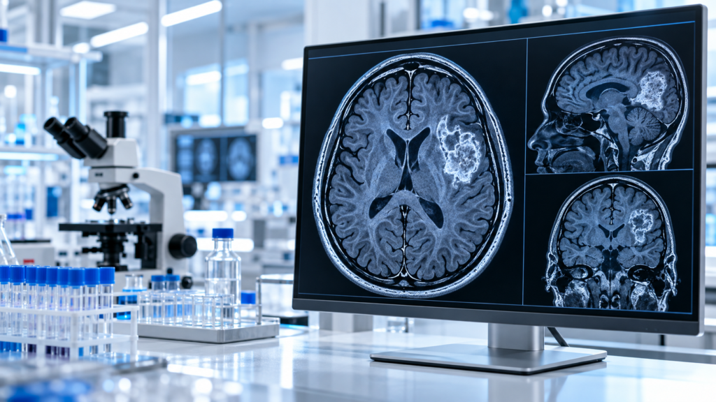
Brain tumor biomarker research has transformed how scientists classify central nervous system tumours. The World Health Organization’s modern classification incorporates molecular information alongside tissue appearance. Markers including IDH mutations, 1p/19q codeletion and MGMT promoter methylation provide biologically important information, although they serve different diagnostic, prognostic and predictive purposes. The US National Cancer Institute documents how these molecular features contribute to contemporary tumour classification and clinical research. [NCI +2]
| MOLECULAR FEATURE | MAIN SCIENTIFIC RELEVANCE | SOURCE |
|---|---|---|
| IDH1/IDH2 mutation | Helps distinguish biologically different glioma categories | NCI central nervous system tumour review |
| 1p/19q codeletion | Required with an IDH mutation for molecular diagnosis of oligodendroglioma | NCI oligodendroglioma guide |
| MGMT promoter methylation | Studied as a prognostic and predictive feature in specific glioma contexts | NCI adult brain tumour review |
| Histone-related molecular alterations | Relevant to classification of selected paediatric gliomas | NCI childhood glioma review |
| Integrated molecular diagnosis | Combines tissue and molecular evidence rather than relying on microscopy alone | NCI childhood glioma review |
Evidence table
The significance of these discoveries is not that scientists have found a single universal biomarker.
It is that researchers now recognize several biologically distinct diseases within categories that once appeared more similar.
This change has important consequences for research design.
When investigators combine molecularly different tumours into a single study population, results may be difficult to interpret.
More precise classification allows investigators to ask more specific questions.
It also encourages better comparison between laboratory models and the actual biological characteristics of tumours.
Brain Cancer Awareness Day and International Brain Tumour Awareness Week provide an appropriate setting to explain this developing science.
The discussion below focuses on biomarkers, laboratory methods and evidence quality.
It does not recommend experimental compounds or claim that research materials prevent or treat cancer.
Table of contents
- Why brain tumor biomarker research matters
- Finding 1: Molecular classification changes disease definitions
- Finding 2: IDH mutations identify important biological differences
- Finding 3: The 1p/19q codeletion helps define oligodendroglioma
- Finding 4: MGMT methylation provides another evidence layer
- Finding 5: Paediatric brain tumours require distinct classification
- Finding 6: Liquid biopsy is an active investigative field
- Finding 7: Laboratory quality determines biomarker reliability
- How biomarker studies should be interpreted
- Research peptides and molecular pathways
- Canadian laboratory research considerations
- Frequently asked questions
- References
Why brain tumor biomarker research matters
Brain tumours are biologically diverse.
Some arise from glial cells or related precursor populations.
Others involve different cell lineages or developmental pathways.
Tumours also differ in anatomical location, molecular characteristics and biological behaviour.
This diversity makes classification unusually important.
Historically, pathologists relied heavily on microscopic appearance to classify tumours.
Histology remains essential.
However, advances in molecular biology have demonstrated that tumours with similar visual appearances may have significantly different genetic characteristics.
Biomarker research examines those distinctions.
The limitations of appearance alone
Microscopy can reveal cell shape, tissue architecture and other features.
These observations provide important diagnostic information.
Yet tissue appearance does not always capture the full biological identity of a tumour.
Two samples may show overlapping morphological features but carry different molecular alterations.
Those differences can affect classification and research interpretation.
The modern approach therefore integrates multiple evidence types.
What a biomarker means
A biomarker is a measurable characteristic associated with a biological process, condition or response.
In oncology, biomarkers can include DNA alterations, gene-expression patterns, protein measurements or epigenetic features.
However, biomarkers have different functions.
A diagnostic biomarker helps identify a condition or subtype.
A prognostic biomarker provides information associated with likely outcomes.
A predictive biomarker provides information about the probability of response to a particular intervention.
These categories overlap in some circumstances but should not be treated as interchangeable.
Why molecular classification improves research
A laboratory finding becomes more informative when investigators know which tumour subtype is being studied.
Molecular classification can reduce the problem of combining biologically different cases.
It also supports more precise comparisons between experiments.
Researchers can investigate whether a proposed mechanism exists consistently within a defined subtype.
That strengthens the interpretation of mechanistic studies.
However, improved classification does not automatically establish an effective intervention.
Classification and treatment development are separate scientific achievements.
Finding 1: Molecular classification has changed the definition of important brain tumours
One of the most consequential advances in this field is the move toward integrated diagnosis.
Modern classification increasingly combines histological and molecular findings.
The WHO’s fifth edition classification of central nervous system tumours introduced a framework that recognizes the biological importance of molecular alterations.
The NCI describes how this framework informs contemporary tumour classification. [cancer.gov]
What integrated diagnosis means
An integrated diagnosis combines information from multiple methods.
A tissue sample may first be examined microscopically.
Pathologists may then assess selected molecular alterations.
The final classification incorporates these findings into a coherent interpretation.
This does not mean every tumour requires the same set of tests.
The appropriate molecular workup depends on the suspected tumour type and applicable diagnostic standards.
Why the change matters
A tumour’s name is not merely descriptive.
It determines how scientists group cases in research datasets.
When classification changes, older clinical trials and published studies may contain tumour populations defined under earlier criteria.
This creates challenges when comparing historical and modern research.
An apparent difference between studies might reflect changes in tumour classification rather than a true biological contradiction.
Researchers need to examine how participants were classified.
Biomarkers and scientific reproducibility
Reproducibility depends on comparing similar biological populations.
If two laboratories study different molecular subtypes but label them identically, their findings may conflict.
Modern classification reduces some of that uncertainty.
It allows researchers to report more precise inclusion criteria.
It also makes negative findings easier to interpret because investigators can evaluate whether results differ between subtypes.
What classification cannot establish
An integrated diagnosis may provide important information about tumour biology.
It does not, by itself, prove that a particular drug or experimental molecule will be effective.
Therapeutic questions require separate evidence.
Researchers must evaluate interventions using appropriate clinical or preclinical methods.
This distinction prevents molecular discoveries from being overstated.
Finding 2: IDH mutations identify biologically important differences
IDH refers to isocitrate dehydrogenase, an enzyme involved in cellular metabolism.
Alterations in IDH1 or IDH2 occur in selected glioma subtypes.
The presence or absence of these alterations can help define biologically distinct groups.
As a result, IDH status is central to biomarker research.
Why metabolism matters
Cells rely on interconnected metabolic pathways.
Enzymes participate in reactions that support energy production, biosynthesis and regulation of cellular processes.
Some IDH mutations alter normal enzyme activity.
These changes can influence metabolite production and downstream molecular regulation.
The resulting biology has helped researchers understand why certain tumour subtypes differ from others.
IDH status and tumour classification
Modern adult diffuse glioma classification uses IDH status as an important molecular feature.
The NCI identifies IDH-related differences in classification and outcomes.
In selected research populations, IDH-mutated tumours have shown different prognostic characteristics from IDH-wildtype tumours. [NCI]
However, the meaning of IDH status depends on tumour type and context.
A single molecular result should not be interpreted independently of histology, additional biomarkers and clinical information.
What observational associations establish
An association between a biomarker and outcome does not automatically establish causation.
For example, a molecular subtype may be associated with longer survival.
That finding does not prove that experimentally altering the biomarker would reproduce the same survival pattern.
It may instead identify a broader biological state involving several interacting pathways.
Researchers therefore study both mechanisms and population-level associations.
Why laboratory models must be carefully selected
A laboratory cell line can provide valuable mechanistic information.
But cell lines may accumulate changes during repeated culture.
They may also fail to reproduce the complexity of a tumour’s original environment.
Researchers should confirm whether models retain the relevant molecular characteristics.
An IDH-related finding in one cell model may not apply to all gliomas.
This is why validation in several complementary models strengthens experimental conclusions.
Finding 3: The 1p/19q codeletion helps define oligodendroglioma
The combined loss of specific portions of chromosomes 1 and 19 is known as the 1p/19q codeletion.
This molecular alteration is central to the modern definition of oligodendroglioma.
According to the NCI, diagnosing oligodendroglioma requires identifying both an IDH mutation and the 1p/19q codeletion. [cancer.gov]
This makes the codeletion a particularly clear example of how molecular research has changed diagnostic classification.
What codeletion means
Human chromosomes contain DNA organized into distinct structures.
Chromosomes have short and long arms.
The designation 1p refers to the short arm of chromosome 1.
The designation 19q refers to the long arm of chromosome 19.
A codeletion involving these regions represents a specific chromosomal alteration.
Researchers can identify such abnormalities using appropriate molecular methods.
Why two findings are required
An IDH mutation alone does not establish oligodendroglioma.
The combination of IDH mutation and 1p/19q codeletion provides the molecular definition.
This illustrates a broader principle.
A single biomarker may be informative without being sufficient for a complete classification.
Some conditions require a particular combination of findings.
Diagnostic testing and assay limitations
Laboratories can investigate chromosomal alterations through several validated molecular approaches.
Different methods have different technical characteristics.
Researchers must consider what genomic regions the assay detects and how results are interpreted.
A technically valid test should distinguish the relevant molecular alteration from other patterns that could produce misleading results.
Quality controls and appropriate interpretation standards are essential.
Implications for historical research
Older publications may describe oligodendroglioma using diagnostic criteria different from those used today.
Some historical cohorts may therefore include cases that would receive different integrated diagnoses under modern classification.
That does not make those studies worthless.
It means comparisons require attention to classification methods.
Researchers should identify which criteria were used when interpreting historical survival or treatment studies.
Finding 4: MGMT promoter methylation adds another layer of molecular evidence
MGMT stands for O6-methylguanine-DNA methyltransferase.
The gene encodes a protein involved in repairing certain forms of DNA damage.
Researchers have studied MGMT promoter methylation because it is associated with clinically relevant biological characteristics in specific glioma contexts.
It is an important component of this research.
DNA repair and tumour biology
Cells contain mechanisms that identify and repair damage to genetic material.
These systems help maintain genomic integrity.
MGMT is one of several components involved in DNA repair.
Changes affecting MGMT expression can alter the activity of this repair pathway.
Promoter methylation is an epigenetic feature that may be associated with reduced gene expression in relevant settings.
However, molecular regulation is complex.
The presence of methylation does not necessarily establish a simple universal relationship between gene expression and biological behaviour.
The distinction between biomarkers
MGMT promoter methylation illustrates why predictive and prognostic terminology must be used carefully.
A prognostic association concerns the relationship between a biological characteristic and outcomes.
A predictive association concerns differential response to a particular intervention.
The NCI describes MGMT promoter status within the evidence base for central nervous system tumours. [NCI]
Interpretation depends on tumour subtype, study population and treatment context.
Laboratory measurement
Methylation studies require methods appropriate to the specific genomic region.
Researchers need to evaluate analytical sensitivity and specificity.
Sample quality also matters.
A tumour tissue specimen may contain both malignant and nonmalignant cells.
The proportion of different cell populations can influence molecular measurements.
Scientists should document assay design and quality-control procedures.
Why cutoffs matter
Some biomarker tests classify results according to a threshold.
For example, a continuous molecular measurement may be converted into categories such as positive or negative.
The selected threshold affects classification.
Different thresholds can produce different proportions of positive cases.
Researchers should therefore explain how cutoffs were established and whether they were validated.
A threshold chosen after examining outcomes in a small dataset may perform less reliably in an independent population.
Finding 5: Paediatric brain tumours require their own molecular frameworks
An important discovery in biomarker research is that paediatric and adult brain tumours cannot always be classified through the same biological assumptions.
Some molecular alterations are associated with particular developmental contexts.
The NCI describes how modern classification distinguishes adult-type diffuse gliomas from several paediatric glioma categories. [cancer.gov]
Why age can reflect biology
Age is not merely a demographic variable.
The nervous system changes throughout development.
Different cellular populations and developmental pathways may influence tumour biology.
Consequently, tumours in children can differ substantially from those more commonly observed in adults.
This means findings from adult tumour studies may not generalize to paediatric disease.
Histone-associated alterations
Researchers have identified specific histone-related molecular alterations associated with selected paediatric central nervous system tumours.
Histones are proteins involved in organizing DNA.
Molecular changes affecting histone biology may influence gene regulation and cellular states.
The biological consequences depend on the exact alteration and tumour context.
A general statement that histone mutations affect all paediatric brain tumours would be inaccurate.
Different classification groups
The modern WHO framework distinguishes several broad categories of paediatric gliomas.
These include paediatric-type diffuse low-grade gliomas and paediatric-type diffuse high-grade gliomas.
Other tumour groups have their own defining features.
The classifications are more complex than a simple division between benign and malignant.
Researchers need to identify the appropriate molecular entity before drawing conclusions.
Study-design implications
Paediatric brain tumour studies can face recruitment challenges because individual molecular subtypes may be uncommon.
Small sample sizes limit statistical precision.
Multi-centre collaboration can improve representation.
However, combining datasets also requires harmonized diagnostic methods.
If participating institutions use different molecular testing standards, comparisons may become unreliable.
Precise classification is therefore especially important in rare-disease research.
Finding 6: Liquid biopsy is an active area of investigation
Traditional molecular analysis often relies on tissue obtained through surgery or biopsy.
Researchers are investigating whether biological material found in bodily fluids could provide additional information.
These approaches are often grouped under the term liquid biopsy.
Liquid biopsy is a developing field within molecular oncology research.
What scientists look for
Investigators may study circulating tumour DNA, cell-free nucleic acids, extracellular vesicles and other molecular signals.
These materials can potentially carry information associated with tumour biology.
However, the detectability of a marker varies.
The biological location of a tumour and the surrounding barriers can influence whether informative material reaches the sampled fluid.
The blood-brain barrier
The blood-brain barrier regulates the movement of many substances between the circulation and the central nervous system.
Its characteristics can complicate blood-based biomarker research for brain tumours.
A low concentration of detectable tumour-associated material may limit assay sensitivity.
The absence of a measurable signal does not necessarily establish that a tumour is absent.
This is an important limitation when evaluating experimental tests.
Other biological sample types
Researchers have also investigated cerebrospinal fluid for molecular analysis in appropriate research and clinical contexts.
Such investigations involve different sampling considerations from ordinary blood tests.
The potential analytical advantages must be evaluated alongside procedural requirements and safety.
An informational research article should not imply that such procedures are suitable for unsupervised use.
Early detection versus monitoring
A biomarker that helps characterize an already diagnosed tumour may not be useful for population screening.
The statistical challenge is different.
When a disease is uncommon, even a test with good laboratory performance may produce many false-positive results in a broad screening population.
Researchers must establish the intended use of each assay.
Possible research objectives include molecular characterization, monitoring and investigation of biological changes.
Each requires different validation.
Why preliminary studies are not enough
Early liquid-biopsy studies may report promising sensitivity or specificity.
However, results can be influenced by sample selection.
A study comparing confirmed advanced tumours with healthy controls may not represent the much harder task of identifying early disease in an unselected population.
Independent validation is necessary.
Prospective studies may also be needed to determine whether using the test improves meaningful outcomes.
Finding 7: Laboratory quality determines whether biomarker results are trustworthy
Molecular discoveries depend on reliable measurements.
This is sometimes overlooked when research results are summarized for general audiences.
A sophisticated molecular assay can still produce misleading findings if samples are mishandled or analytical methods are inadequate.
Quality assurance is therefore fundamental to this research.
Pre-analytical variables
Pre-analytical factors arise before the measurement itself.
They may include specimen collection, preservation, transportation and storage.
For tissue specimens, processing methods can influence nucleic-acid quality.
Researchers should document relevant handling conditions.
These details may explain differences between laboratories.
Analytical validity
Analytical validity concerns whether an assay accurately measures its intended target.
Relevant characteristics include accuracy, reproducibility, sensitivity and specificity.
A test may detect a molecular alteration reliably in one specimen type but perform differently in another.
Researchers must establish validity within the intended context.
Reference materials and controls
Appropriate controls help detect technical problems.
Positive controls can demonstrate that an assay can identify the intended target.
Negative controls can help reveal contamination or nonspecific signals.
Reference materials may support consistency across measurement runs.
However, quality assurance must be designed around the actual method.
There is no universal control strategy suitable for every biomarker assay.
Interlaboratory reproducibility
Different laboratories may use different platforms.
Comparability requires more than reporting the same biomarker name.
Methods, analytical thresholds and reporting conventions may differ.
Interlaboratory studies can reveal systematic variation.
Standardization helps researchers determine whether apparent biological differences are genuine or partly methodological.
The importance of transparency
Scientific publications should describe their analytical procedures.
They should disclose the limitations of the methods used.
When commercial assays or proprietary technologies are involved, conflicts of interest and funding should be reported.
The objective is not to reject industry research automatically.
It is to allow readers to evaluate the evidence appropriately.
Independent replication
A result that appears promising in one dataset may not reproduce elsewhere.
Replication is especially important for exploratory biomarker discovery.
Large datasets can generate statistically significant associations that lack practical value.
Researchers should distinguish hypothesis-generating discoveries from independently validated biomarkers.
That distinction protects the field from premature claims.
How to interpret a brain tumour biomarker study
A useful evidence review begins with the study design.
Was the investigation conducted in cultured cells, animal models, stored human tissue or a prospective patient cohort?
These are different evidence categories.
A finding at one level should not automatically be transferred to another.
Examine the population
The study should describe how tumour cases were classified.
Were molecular criteria applied consistently?
Did the researchers include adult and paediatric cases?
Were different tumour grades combined?
These decisions influence interpretation.
Examine the biomarker method
The assay should be appropriate for the molecular feature being studied.
Investigators should explain how they handled uncertain results.
They should also report whether laboratory personnel were blinded to relevant clinical outcomes when appropriate.
Blinding can reduce certain sources of bias.
Examine the endpoint
A study might evaluate diagnostic accuracy.
Another might examine progression-free survival.
A third might investigate a biochemical mechanism.
The results should be interpreted according to the actual endpoint.
A correlation with a laboratory measurement is not evidence of improved survival.
Examine the sample size
Small studies can be useful for identifying hypotheses.
However, estimates from small samples often have considerable uncertainty.
Subgroup analyses can become especially unstable.
A study involving dozens of cases should not be described as definitively establishing a universal biological relationship.
Examine the confidence intervals
Point estimates provide incomplete information.
Confidence intervals help describe statistical uncertainty.
Wide intervals may indicate that the true effect could differ substantially from the reported estimate.
A statistically significant result can still have limited practical importance.
Conversely, an inconclusive result from a small study does not necessarily demonstrate that no biological relationship exists.
Examine the comparator
A biomarker should be evaluated against an appropriate standard.
If a new assay is proposed as an improvement, researchers should establish what it improves upon.
An assay that merely reproduces information already available through established methods may offer limited additional utility.
The practical value of the test depends on its intended application.
Research peptides, signalling pathways and molecular oncology
Peptides and proteins participate in many biological signalling systems.
Researchers investigate these systems to understand cellular behaviour.
However, their relevance to molecular oncology requires precise interpretation.
Studying a pathway is not the same as establishing a therapy
A laboratory experiment might show that a peptide influences a signalling pathway in a controlled model.
That result may help scientists investigate a biological mechanism.
It does not establish clinical efficacy.
It also does not establish appropriate human exposure, safety or regulatory authorization.
The distinction is particularly important in oncology, where biological pathways can have different effects across tissues and tumour subtypes.
Model-dependent effects
The same molecular pathway may behave differently depending on cellular context.
Genetic alterations can change signalling relationships.
Tumour microenvironments also contain multiple interacting cell populations.
Consequently, an experimental effect observed in one model may not appear in another.
Researchers should avoid generalizing from isolated systems.
Compound identity and analytical quality
For legitimate laboratory research, investigators need to establish the identity and relevant analytical characteristics of research materials.
Methods such as chromatography and mass spectrometry can support analytical characterization.
However, chemical purity does not demonstrate biological efficacy or human safety.
These are separate scientific questions.
Readers exploring basic analytical terminology can consult the Northern Peptides Educational Resources.
The site’s Research FAQ provides additional background about its laboratory-research focus.
These internal resources are general scientific references, not oncology treatment recommendations.
Why the distinction protects research quality
Overstated claims can damage trust in legitimate science.
A promising cellular observation may deserve further investigation.
But describing it as proof of a human treatment bypasses essential experimental stages.
Responsible research communication preserves the difference between a hypothesis and an established result.
The Canadian research perspective
Canada has academic institutions, hospitals and research centres contributing to molecular biology and oncology.
Researchers operate within institutional ethics and safety requirements appropriate to their work.
Molecular studies involving human tissue or identifiable information require particular attention to ethical approval, consent and privacy.
Data protection
Genomic information can be highly sensitive.
Researchers must manage access appropriately.
De-identification procedures can reduce certain risks, but genomic data require careful governance.
Data sharing should occur under applicable institutional and legal frameworks.
Collaborative research
Rare molecular tumour subtypes can be difficult to study at a single centre.
Collaboration across institutions may increase sample sizes.
However, pooled datasets need consistent definitions.
Differences in assay methods, diagnostic criteria and follow-up practices can create misleading comparisons.
Harmonization is therefore an important component of collaborative research.
Open scientific questions
Several questions remain unresolved.
Which molecular features most reliably distinguish clinically meaningful subtypes?
How can molecular assays become more reproducible?
Which biomarkers provide information beyond established classification systems?
Can minimally invasive methods achieve adequate sensitivity for defined applications?
How can investigators validate results across populations and technologies?
These questions show why biomarker research remains an active scientific field.
What the seven discoveries mean together
The evolution of molecular classification represents genuine scientific progress.
Researchers now understand that central nervous system tumours cannot be classified solely through a single visual characteristic.
Genetic, chromosomal and epigenetic findings contribute important information.
IDH status demonstrates the relevance of altered cellular metabolism.
The 1p/19q codeletion illustrates how combinations of biomarkers can define a tumour entity.
MGMT promoter methylation highlights relationships between molecular regulation and clinical research.
Paediatric classification demonstrates that tumour biology differs across developmental contexts.
Liquid-biopsy investigations are testing possible new measurement approaches.
And laboratory quality assurance remains essential for every discovery.
The greatest value of brain tumor biomarker research lies in making biological questions more precise.
That precision can improve study design and the interpretation of evidence.
It does not eliminate uncertainty.
Nor does it justify unsupported therapeutic claims.
The most credible scientific progress combines promising discoveries with careful validation.
Frequently asked questions
1. What is brain tumor biomarker research?
Brain tumor biomarker research investigates measurable molecular and biological characteristics associated with central nervous system tumours. These may contribute to classification, prognosis or research into biological mechanisms.
2. Why is IDH mutation status important?
IDH alterations help distinguish biologically different glioma categories. Their interpretation depends on the specific tumour type and other molecular and pathological findings.
3. What does the 1p/19q codeletion indicate?
The 1p/19q codeletion is a chromosomal alteration used with an IDH mutation to define oligodendroglioma under modern molecular classification.
4. Can liquid biopsy replace tissue-based molecular diagnosis?
Liquid biopsy is an active research area. Its usefulness depends on the application, biological sample and analytical validation. Experimental findings do not establish it as a universal replacement for tissue-based diagnosis.
5. Do research peptides treat brain tumours?
Laboratory findings involving peptides or signalling pathways do not establish that experimental peptide materials treat brain tumours. Such claims would require appropriate clinical evidence and regulatory assessment. This article makes no therapeutic recommendation.
References
- National Cancer Institute. Central Nervous System Tumors Treatment (PDQ).
- National Cancer Institute. Oligodendroglioma and Other IDH-Mutated Tumors.
- National Cancer Institute. Childhood Astrocytomas and Other Gliomas Treatment (PDQ).
- National Cancer Institute. Childhood Cancer Genomics (PDQ).
- Canadian Cancer Society. Canadian Cancer Statistics 2025.
For laboratory research use only. Not for human consumption.